
When Pregnant, When Do You Get Your First Ultrasound?
A mother’s joy begins when new life is stirring inside, when a tiny heartbeat is heard for the first time, and a playful kick reminds her that
Are you ready to witness the miraculous moments of your baby’s early development? Imagine seeing their tiny heart beating for the first time, confirming the joyous reality of your pregnancy.
The 8-week ultrasound brings you closer to your little one, allowing you to glimpse their growing limbs, developing organs, and the promise of new life.
At Choices Women’s Clinic, we understand the significance of the 8-week ultrasound during your pregnancy journey.
As an expectant mother, you may have questions and concerns about what to expect during this crucial milestone. This is an exciting milestone in your pregnancy journey.
This article will guide you through what to expect during your 8-week ultrasound and provide essential information to ease any anxieties. Let’s get started.

The 8-week ultrasound scan holds significant importance in your pregnancy journey. It serves multiple purposes, helping healthcare professionals confirm your pregnancy, estimate your due date accurately, and assess the well-being of your developing baby.
At Choices Women’s Clinic, our dedicated team of experts is committed to providing comprehensive care during this pivotal stage of your pregnancy.
During the ultrasound at 8 weeks, several important things happen as healthcare professionals gather valuable information about your pregnancy. Here’s what you can expect during this significant stage:
The primary purpose of the 8th pregnancy week ultrasound is to confirm the presence of a viable pregnancy. The sonographer will carefully examine your uterus and look for the gestational sac, a fluid-filled structure surrounding the developing embryo.
The sonographer will measure the crown-rump length (CRL) of your baby. This measurement helps estimate your due date accurately and determines the stage of fetal development. At 8 weeks, your baby is typically about the size of a raspberry or nearly the same size as any small berry.
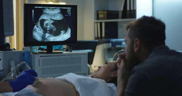
The ultrasound will provide a clear image of the gestational sac, the early structure that houses your developing baby. The sac appears as a black circle on the ultrasound screen filled with amniotic fluid.
One of the most incredible moments during the 8-week ultrasound is hearing and seeing your baby’s heartbeat. The sonographer will use Doppler technology or other methods to detect the rhythmic beats of your baby’s tiny heart. A strong and regular heartbeat is an encouraging sign of a healthy pregnancy.
The yolk sac, another essential structure, will be visible on the ultrasound. It provides vital nutrients to your developing baby until the placenta takes over this role later in pregnancy.
The sonographer may also examine your uterus and ovaries to ensure no abnormalities or complications could affect your pregnancy.
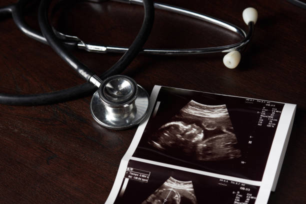
During the 8-week ultrasound, the sonographer will capture images and measurements of these critical aspects. This information helps healthcare providers monitor the progress of your pregnancy, estimate your due date, and ensure that everything is developing as expected.
It’s important to note,
The 8-week ultrasound is a non-invasive and safe procedure for you and your baby. It provides valuable insights into the early stages of fetal development, allowing you to visualize your little one’s growth and strengthening the emotional connection with your pregnancy.
At Choices Women’s Clinic, our state-of-the-art ultrasound technology allows you to witness incredible details during your 8-week ultrasound. Though your baby is still in the early stages of development, the ultrasound images will unveil remarkable features, including:
As you witness your baby’s tiny heart beating rhythmically on the screen, prepare to be amazed. The sonographer will measure the heartbeat rate, providing an important indicator of your baby’s well-being.
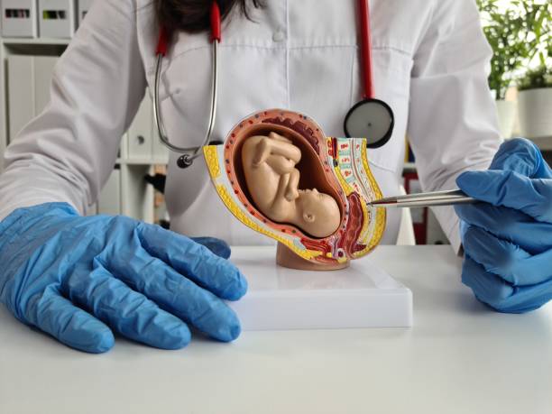
During the 8-week ultrasound, you’ll have the opportunity to see the beginnings of your baby’s limbs, limb buds, and arm and leg buds. A Baby’s head will appear larger in proportion to the body, which is crucial in housing your baby’s developing brain.
Although still in the early stages of the first trimester, the ultrasound may reveal the presence of basic internal organs, such as the stomach and kidneys, as your baby’s body takes shape.
The amniotic fluid surrounding your baby will be visible on the ultrasound images creating a serene and protective environment for their growth and development. This cushion fluid ensures your baby is safe and comfortable throughout pregnancy.
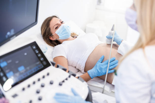
Regarding early ultrasound during pregnancy, there are two common methods: transvaginal and abdominal ultrasound. Each method has its benefits and considerations. Let’s explore the differences between these two types of ultrasounds:
A transvaginal ultrasound involves inserting a specially designed wand or probes into the vagina to obtain images of the pelvic organs and the developing fetus.
Here are some key points about transvaginal ultrasounds:
Before the early ultrasounds procedure, you will be asked to empty your bladder during a transvaginal ultrasound. The ultrasound technician or healthcare provider will insert the probe into the vagina, which may cause mild discomfort but is generally well-tolerated.
Transvaginal ultrasounds provide clearer and more detailed images in an early pregnancy scan. The proximity of the probe to the uterus allows for a closer view of the developing embryo and better visualization of structures.
Transvaginal ultrasounds are particularly useful in the early stages of pregnancy, typically up to around 10 weeks. They can detect pregnancies earlier than abdominal ultrasounds, especially in cases where a clear image cannot be obtained through the abdomen.
Transvaginal ultrasounds also allow for a more comprehensive assessment of the pelvic organs, including the uterus, ovaries, and surrounding structures. This can be beneficial for evaluating any potential issues or abnormalities.
Some individuals may find transvaginal ultrasounds more invasive or uncomfortable than abdominal ultrasounds. However, the procedure is relatively quick and is performed in a private, intimate setting.

An abdominal ultrasound is a more common type of ultrasound that involves placing a transducer on the surface of the abdomen to capture images of the pelvic organs and the developing fetus.
Here’s what you need to know about abdominal ultrasounds:
For an abdominal ultrasound, you will be asked to have a full bladder to help improve image quality. The transducer is moved gently over the abdomen, transmitting sound waves and creating images on a monitor.
Abdominal ultrasounds provide a broader view of the pelvic area, including the uterus, ovaries, and surrounding structures. They can be used to assess the overall health of these organs and detect any abnormalities.
Abdominal ultrasounds are commonly performed throughout pregnancy, including the later stages. As the baby grows and the uterus rises higher in the abdomen, abdominal ultrasounds more effectively capture images.
Abdominal ultrasounds are non-invasive and generally painless. They do not require any insertion into the body and are typically well-tolerated by most individuals.
In early pregnancy, particularly before 10 weeks, abdominal ultrasounds may not provide as clear and detailed images as transvaginal ultrasounds. The distance between the transducer and the developing embryo can sometimes limit visibility.
Know that,
The choice between transvaginal and abdominal ultrasound depends on various factors, including the stage of pregnancy, individual circumstances, and the preferences of both the healthcare provider and the patient.
Your healthcare provider will recommend the most appropriate type of ultrasound based on your specific needs.
In some cases, during an ultrasound, it may not be possible to see the baby or detect a heartbeat. This can understandably be concerning for expectant parents. Here are a few possible reasons why this might occur:
If the ultrasound is performed very early in the pregnancy, typically before 6-7 weeks, it may be challenging to visualize the baby or detect a heartbeat. The embryo is still very small at this early stage, and it may take a little more time to be visible on the ultrasound.
Sometimes, the estimated gestational age based on the last menstrual period may not align with the actual stage of pregnancy. If the ultrasound is performed too early or too late based on the estimated dates, it may result in difficulties visualizing the baby or the heartbeat.
The baby’s position within the uterus can also affect visibility during an ultrasound. If the baby is positioned in a way that makes it challenging to obtain clear images, it may be difficult to see them or detect a heartbeat. The healthcare provider may recommend a follow-up ultrasound or alternative imaging techniques.
Unfortunately, the inability to see the baby or detect a heartbeat may sometimes indicate a miscarriage or pregnancy loss. This can be a devastating and emotional experience for expectant parents.
Also, check for ectopic pregnancy issues. If there are concerns about a possible miscarriage, additional diagnostic tests and follow-up appointments may be necessary to confirm the situation.
![]()
At 8 weeks pregnant, you are entering an exciting phase of your pregnancy journey. To help you stay organized and ensure you’re taking care of yourself and your growing baby, here’s a pregnancy checklist specifically tailored to this stage:
Regular prenatal care is essential for monitoring your and your baby’s health and development. If you haven’t already, schedule your first prenatal appointment with your healthcare provider. They will conduct tests, review your medical history, and provide important guidance.
Prenatal vitamins provide essential nutrients to support your baby’s growth and development. Talk to your healthcare provider about the recommended prenatal vitamins, including folic acid, iron, and other necessary supplements.
Inform your healthcare provider about any medications or supplements you are currently taking. They will assess their safety during pregnancy and make any necessary adjustments to ensure your and your baby’s well-being.
Focus on a balanced diet that includes a variety of fruits, vegetables, whole grains, lean proteins, and healthy fats. Opt for nutrient-rich foods to support your baby’s growth. Avoid consuming raw or undercooked meats, fish with high mercury content, unpasteurized dairy products, and excessive caffeine.
Drink plenty of water daily to stay hydrated and support your overall well-being. Proper hydration is important for maintaining amniotic fluid levels and aiding in digestion.
Engage in moderate exercise, as approved by your healthcare provider. Walking, swimming, and prenatal yoga can help maintain your fitness and prepare your body for labor. Avoid high-impact or strenuous exercises without medical guidance.
Take the time to educate yourself about pregnancy, childbirth, and parenting. Attend prenatal classes or workshops to gather information and prepare yourself for the upcoming journey.
Pregnancy can bring about a range of emotions. Take care of your mental and emotional health by seeking support from your partner, loved ones, or a counselor.
Also, seek your doctor if you feel any imbalance in the pregnancy hormones. To reduce stress and anxiety, practice relaxation techniques like deep breathing and meditation.
It is crucial to avoid alcohol, tobacco, and recreational drugs during pregnancy, as they can harm your baby’s development. If you need assistance quitting smoking or managing substance use, seek support from your healthcare provider.
Read up on the common physical and emotional changes that occur during pregnancy. Familiarize yourself with symptoms like morning sickness, breast tenderness, fatigue, and mood swings to better understand what to expect.
At Choices Women’s Clinic, we are dedicated to providing you with exceptional care and guidance during your pregnancy journey. The 8-week ultrasound is a significant milestone that offers a glimpse into the remarkable progress of your baby’s development.
From witnessing their tiny heartbeat to observing the early signs of their limbs and organs, this experience is filled with wonder and excitement. Contact Choices Women’s Clinic today to schedule your 8-week ultrasound and embark on this incredible journey of parenthood with confidence and peace of mind.
A mother’s joy begins when new life is stirring inside, when a tiny heartbeat is heard for the first time, and a playful kick reminds her that

Carrying a baby is the most rewarding experience a woman can enjoy. But having a baby also comes with its challenges, including pregnancy insomnia. Pregnancy

At Choices Women’s Clinic, we understand the challenges that unexpected pregnancies can bring, and we are committed to providing a supportive environment for individuals facing

Boundaries are the psychological and emotional lines we draw between ourselves and others to ensure our safety, well-being, and happiness. Healthy sexual boundaries in a relationship

People usually use the terms STI and STD interchangeably, but there is a difference between them. STI stands for sexually transmitted infection, while STD stands

It seems like from the moment you learn about sex at a young age, the pressure to have sex is on. Whether you are single,