A mother’s joy begins when new life is stirring inside, when a tiny heartbeat is heard for the first time, and a playful kick reminds her that she is never alone. Ultrasound is the technology that allows us to peek inside and see the miracle of life in its earliest stages.
Most women will receive at least one ultrasound during their pregnancy. An early 6 week ultrasound can be performed as soon as six weeks after your last menstrual period (LMP), though it isn’t always necessary.
A dating ultrasound may then be scheduled to confirm the age of the fetus. The first-trimester screening ultrasound, typically performed between 10 and 14 weeks, a 13 week ultrasound is usually done in stock for potential problems with the baby’s development or anatomy.
Choices Women’s Clinic offers a 3D/4D ultrasound, usually performed around 20 weeks gestation, to help parents bond with their unborn child. This blog post will provide more information on when to expect an ultrasound and how CWC can assist you throughout the process. Let’s begin.
What is Ultrasound | A General Overview

Ultrasound is a non-invasive imaging technology that uses high-frequency sound waves to create images of the inside of a woman’s body.
These images can be used to show the development and growth of the fetus throughout pregnancy, determine the baby’s age, check for any potential abnormalities or genetic conditions, and assess the position of the placenta.
Why Is A First Fetal Ultrasound Necessary During Pregnancy?

Ultrasound is among the few ways your pregnancy care provider can see and hear your baby, providing valuable insights into your pregnancy’s progression. It helps determine gestational age, tracks proper growth and development, and identifies potential complications.
Depending on what your provider is looking for, ultrasounds may occur anytime during pregnancy. Not all women have a first-trimester ultrasound.
Having just one ultrasound during pregnancy is standard; a mid-pregnancy transabdominal ultrasound between 18 and 22 weeks. This is sometimes called an anatomy ultrasound because it evaluates your baby’s anatomy.
First Trimester Ultrasound | What You Need To Know
The first-trimester ultrasound is usually performed between 6 and 13 weeks of pregnancy. Although not all pregnancies undergo a first-trimester ultrasound, healthcare providers may use them to determine viability, date the pregnancy, or diagnose suspected complications.
Transvaginal ultrasound also makes it easier to diagnose early pregnancy problems, such as a miscarriage or a molar or ectopic pregnancy.
Early Pregnancy (6–8 Weeks)

Your first pregnancy ultrasound, also known as a fetal ultrasound or sonogram, could occur as early as 6 weeks after your last menstrual period (LMP). Early ultrasounds may be done to determine if the pregnancy is viable and located in the uterus, as well as to estimate the age of the fetus.
The first-trimester ultrasound can also detect a heartbeat and assess a baby’s anatomy. An ultrasound is typically performed transvaginally during early pregnancy, which offers the clearest vision of the uterus and embryo at this stage.
In this instance, the healthcare provider will insert a thin wand-like transducer probe into your vagina, which emits high-frequency sound waves through your uterus.
These waves reflect off the fetus and are transmitted back to a machine, which converts them into a black-and-white image of your uterus.
Nuchal Translucency Ultrasound (10–13 Weeks)
The nuchal translucency ultrasound is typically performed between 10 and 13 weeks.
This exam measures the fluid thickness beneath your baby’s neck, which can help identify pregnancies at risk for chromosomal abnormalities, such as Down syndrome or trisomy 18.
The healthcare provider may also use this test to confirm fetal age and assess the anatomy of your baby’s brain, heart, spine, kidneys, and other organs.
Results Of Ultrasound
During pregnancy, an ultrasound can detect if your pregnancy is in your uterus and not ectopic.
It can also determine the number of babies you’re carrying, screen for genetic disorders, identify problems with your placenta, uterus, or ovaries, and assess viability by detecting a heartbeat. Typically, you’ll see your baby’s heartbeat if you’re at least 6 weeks pregnant.
Second Trimester Ultrasound
The second-trimester ultrasound is typically performed between 18 and 22 weeks of pregnancy.
The second trimester is the most common time for a routine prenatal ultrasound. The second Trimester Ultrasound is done through an anatomy scan.
Anatomy Scan | Level 2 Ultrasound
The second-trimester anatomy scan is often called Level 2 ultrasound. An anatomy scan, a thorough scan of your baby’s developing body and organs, is offered to every pregnant person.
The second-trimester ultrasound can also help determine the sex of the baby if desired. Additionally, this ultrasound can identify any complications, such as placental abnormalities or intrauterine growth restriction.
Results
During this ultrasound, the healthcare provider will check the baby’s organs, limbs, and facial features, as well as measure the size and growth rate of the fetus.
During a prenatal check-up, your healthcare provider will conduct several tests to assess your baby’s health and development.
These tests include checking for physical abnormalities, measuring the size of your baby, assessing the amount of amniotic fluid in your uterus, and determining whether you’re carrying one baby or multiple.
Additionally, your healthcare provider will check your baby’s organs, limbs, facial features, heartbeat, position, and movement. They will also measure your cervical length and check the location of your placenta.
Finally, they will try to determine your baby’s sex, providing you with a comprehensive evaluation of your baby’s health and well-being.
Third Trimester Ultrasound

The third-trimester ultrasound is typically performed between 28 and 40 weeks of pregnancy. An ultrasound is often performed on asymptomatic or symptomatic patients, but not always necessary in the third trimester.
However, if your pregnancy is high-risk, it may be recommended. During third trimester ultrasound, they may also use Doppler flow studies to measure blood flow or amniocentesis to determine any genetic problems with the fetus.
Doppler Ultrasound
A Doppler ultrasound is typically performed later in pregnancy to examine how your baby’s blood flows through its blood vessels. This type of ultrasound is used to assess your baby’s health and ensure proper blood circulation.
Fetal Echocardiogram
This ultrasound examines your baby’s heart size, shape, function, and structure. Your healthcare provider may recommend it if your baby is suspected of having a congenital heart condition or if you have a child with a heart condition history.
Additionally, your provider may suggest a closer look at the heart if you have certain health conditions.
Results
The results of a third-trimester ultrasound can provide some insight
The third-trimester ultrasound helps your healthcare provider track the baby’s growth, assess the amount of amniotic fluid, check for any potential abnormalities or problems with the placenta, and confirm that your baby is in a head-down position, ready to be born.
Pregnancy First Ultrasound | The Process

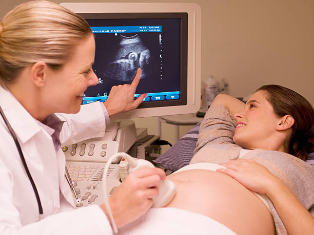
During a pregnancy ultrasound, the healthcare provider will apply a special gel to your abdomen and use a transducer, a wand-like probe, to take images of your baby.
The first step in obtaining an ultrasound is for the healthcare provider to explain what he or she is looking for and any necessary preparations before the ultrasound.
The actual exam itself usually takes between 10 and 45 minutes. During the ultrasound, you can expect to hear your baby’s heartbeat and watch your baby move around inside of you.
Depending on what the healthcare provider is looking for in the ultrasound, he or she may take measurements of various parts of the fetus. Afterward, you’ll be able to see a picture or video clip of your baby.
During Transvaginal Ultrasound
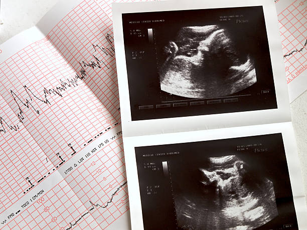
The transducer emits sound waves, which bounce back and create images on a video monitor, allowing you to see your baby. Bones appear white, fluid is black, and soft tissue organs display various shades of gray.
The sonographer records important information and captures images or videos for your healthcare provider to evaluate. You can observe the process on the screen if you wish. Your provider will discuss the findings with you.
During Transabdominal Ultrasound
A sonographer glides a handheld device, roughly the size of a bar of soap, over your stomach and transmits sound waves. A computer then translates the echoes into images displayed on a video monitor, revealing your baby in real-time.
White areas represent bone, black indicates fluid, and soft tissue organs appear as shades of gray. The sonographer records your baby’s measurements and captures still images or videos for your provider to interpret. You can observe the process on the screen if you choose.
Your provider will discuss the results with you, and you may request printed or digital copies of the images to take home.

For complete guidance, please book an appointment at Choices Women’s Clinic. Our experienced healthcare providers will provide the best care possible during your pregnancy journey. Let us help you enjoy this miraculous experience!
At Choices Women’s Clinic, we aim to provide you with the best care possible as you go through this incredible experience of bringing a new life into the world.
Are Ultrasounds Safe | If Yes, How Many?

Yes, ultrasound is considered safe and has been used since the 1960s. While no known adverse effects are associated with ultrasound, following your healthcare provider’s instructions closely is essential.
Depending on your pregnancy and health history, you may need only one or two ultrasounds during your pregnancy. Your healthcare provider will be able to let you know how many ultrasounds are necessary for your personal health and that of the baby.
While pregnancy ultrasounds are generally considered safe in medical environments, improper use can result in tissue heating or the production of bubbles. It is crucial to perform ultrasounds correctly to avoid these risks.
The long-term effects of heated tissues or cavitation from ultrasounds are uncertain, particularly when they are not medically necessary.
Consequently, the FDA recommends that individuals use ultrasound scans judiciously – only when medically required, based on a prescription, and administered by a properly-trained healthcare professional.
Overall, ultrasound is an invaluable tool in determining the health and development of your baby throughout pregnancy.
How Long Does An Ultrasound Take?
Ultrasound exams typically take between 10 and 45 minutes, depending on the reason for the exam. During an ultrasound scan, your healthcare provider will move a transducer wand over your abdomen or sometimes insert it into your vagina to capture images of your baby.
The sonographer will then evaluate these images and record important information for your provider to interpret. After the exam, your provider will discuss the results with you.
If you opt to receive printed or digital copies of images from your ultrasound, this will likely add 10-15 minutes to the total time.
Wrap Up
Ultrasound is a safe, non-invasive procedure to examine your baby’s development during pregnancy. The number of ultrasounds you receive depends on your health history and that of your baby; most people require one or two scans throughout their pregnancy.
It is better to have the first ultrasound in the first trimester for better accuracy and be aware of the baby’s development. However, having an ultrasound scan in the second trimester is normal. If you require ultrasound scan services, Choices Women’s Clinic is here to provide the best care.
With state-of-the-art ultrasound technology and experienced, highly qualified medical practitioners, Choices Women’s Clinic is here to help you every step of the way. Contact us today for more information on ultrasound scans!
Frequently Asked Questions
Is 7 weeks too early for an ultrasound?
They look at your baby’s measurements, which are very predictable in the first 8-11 weeks. An ultrasound up to 12 weeks should be able to predict your gestation accurately within 3-5 days. 7 weeks is definitely not too early to see your baby, but the image might differ from what you imagined.
How do I prepare for my first ultrasound?
No special preparation is needed for an ultrasound. However, some pregnancy care providers may require you to have a full bladder and avoid using the restroom before the test. This is to ensure a better view of your baby on the ultrasound.